
It has been reported that LPS can heal wounds and suppress inflammation.
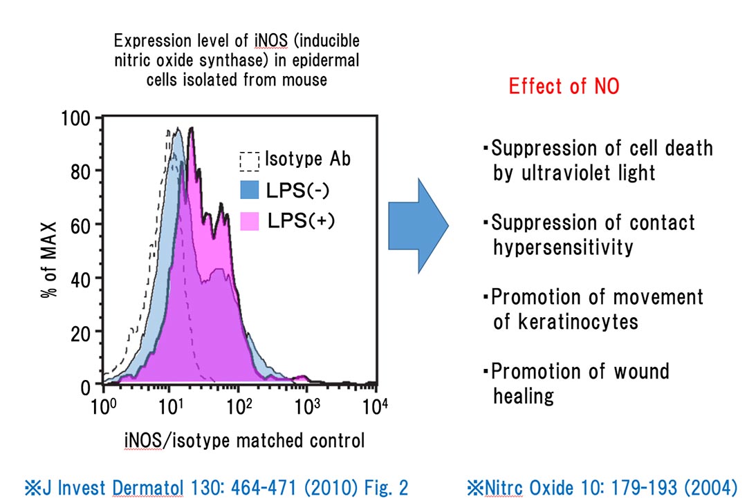
When stimulated by LPS, keratinocytes release NO, which is effective in suppressing cell death associated with ultraviolet radiation, suppressing contact hypersensitivity, and promoting the mobility of keratinocytes (*1). Thus, NO produced by keratinocyte with LPS accelerates wound healing.
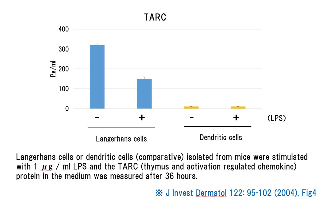
In Langerhans cells on the epidermis, LPS stimulation reduces the expression of inflammatory chemokines called TARC (*2).
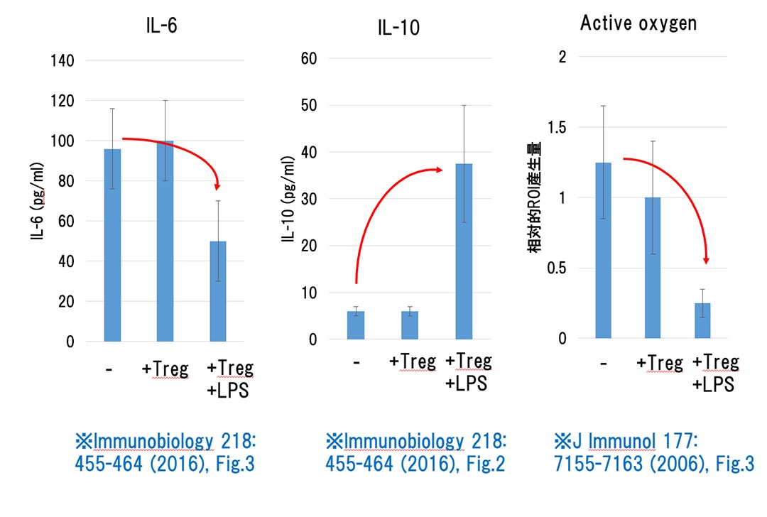
Furthermore, regulatory T (Treg) cells, which mediate inflammation in a suppressive manner, are activated by LPS in the epidermis. The activated Treg cells in turn suppress the inflammatory effects of neutrophils, specifically reducing IL-6 expression, an inflammatory cytokine produced by neutrophils, while enhancing IL-10 expression, an anti-inflammatory cytokine (*3). The activated Treg cells also reduce the quantity of oxygen generated by neutrophils (*4). Of the T cells in the epidermis, the inflammation-promoting T cell does not respond to LPS (*5).
Thus, LPS in the epidermis is effective in preventing skin roughness and relieving inflammation.
(*1) Nitric oxide function in the skin, Nitrc Oxide 10: 179-193 (2004)
(*2) Differential Expression and Function of Toll-like Receptors in Langerhans Cells: Comparison with Splenic Dendritic Cells, J Invest Dermatol 122: 95-102 (2004), Fig4
(*3) Apoptotic epithelial cells control the abundance of Treg cells at barrier surfaces, Nature Immunobiology 218: 455-464 (2016), Fig.2, 3
(*4) Lipopolysaccharide-Activated CD4+CD25+T Regulatory Cells Inhibit Neutrophil Function and Promote Their Apoptosis and Death, J Immunol 177: 7155-7163 (2006), Fig.3
(*5) Regulatory T Cells Selectively Express Toll-like Receptors and Are Activated by Lipopolysaccharide, J. Exp Med. 197: 403-411 (2003) Fig.1

DynaxT bldg. 2F, 2217-6
Hayashi-cho, Takamatsu-shi,
Kagawa-ken,
761-0301 Japan
TEL : +81-87-867-7712
FAX : +81-87-867-7737

Your personal information on this site is protected by SSL.
![]()